Understanding the timing of heart development in Down syndrome
Down syndrome is the most common birth defect syndrome, caused by an extra copy of chromosome 21 (sometimes called trisomy 21). Children with Down syndrome are born with a variety of organ defects, including a high rate of congenital heart disease (CHD). Researchers have struggled to understand how an extra copy of chromosome 21 causes abnormal organ development in children with Down syndrome. Structural issues with the heart and brain in Down syndrome are linked to smaller organs or reduced growth, but it's not well understood how trisomy 21 causes these developmental deficits, leading to CHD and intellectual disability.
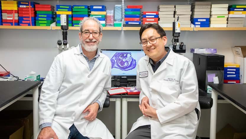
Ivan Moskowitz, MD, PhD, Professor of Pediatrics, Human Genetics, and Pathology at the University of Chicago, has spent his career studying heart development and CHD, focusing on how stem cells differentiate and develop into mature cells that form organs and tissues. Over the years, his lab has shown that a key consideration is not so much how cells differentiate, but when.
Stem cells that are in transition towards their final differentiated cell type, such as heart muscle cells (cardiomyocytes) or neurons, are called progenitor cells. The heart is formed by two different groups of progenitors. The first differentiates into muscle cells early and forms a primitive heart tube. The second waits nearby and differentiates later into muscle cells that form specific structures in the heart required for lung circulation in mammals.
Moskowitz and his lab have shown that if the second group’s timing is off, for example by differentiating too early, the heart will not form properly, resulting in CHD. Furthermore, they have found that a communication process between cells called Hedgehog signaling serves as the timer for differentiation of progenitors as the organ undergoes formation. The pathway was originally discovered in Drosophila fruit flies in the 1970s, and is required for proper patterning of body segments of the flies from the egg. Flies with a mutant version of one of the Hedgehog signaling genes developed a dense “hairy” cuticle and stubby legs that caused them to look like mini-Hedgehogs — thus the name “Hedgehog signaling.”
The Hedgehog pathway has been studied extensively by many labs around the world and has since it was linked to the development of many organs as well as the growth of some tumors. But, remarkably, many unanswered questions remain.
“Despite the fact that there are thousands of papers that have been published on Hedgehog signaling, how it controls differentiation timing to affect organ development or promote cancer isn't well understood at a mechanistic level,” Moskowitz said,
Moskowitz’s lab was led to Hedgehog signaling over 10 years ago, from a study of gene mutations that caused congenital heart defects modeled in mice. They identified mutations that diminished Hedgehog signaling in several different mouse lines, all of which presented with CHD. Their more recent mechanistic studies showed that Hedgehog signaling is only active in the delayed second group of heart cell progenitors, not the mature heart itself. Using a variety of genetic tools, they found that when they switched off Hedgehog signaling in progenitor cells, they turned into heart muscle too soon, or “precociously.”
“That was the first hint suggesting that Hedgehog signaling may be a developmental timer for heart development," Moskowitz explains. "We have subsequently done many experiments that have implicated Hedgehog signaling as a developmental timing switch that is active in progenitors to prevent their differentiation. When Hedgehog signaling is inappropriately diminished, the progenitors differentiate too soon, affecting organ formation.”
This timing can make all the difference in forming the heart properly. If the second group of progenitor cells differentiates too soon, it does not contribute properly to the complete, mature heart. The result is CHD, including septal defects, or holes between heart chambers that require surgery when a patient is a newborn.
Moskowitz and his lab now hope to contribute more to this body of research in the context of Down Syndrome, with the support of an $8 million grant from the National Institute of Child Health and Human Development (NICHD), a division of the National Institutes of Health. In the funded study, they will investigate the possibility that altered Hedgehog signaling contributes to CHD and other organ developmental abnormalities in Down syndrome.
Moskowitz’s team noted that removing Hedgehog signaling from the mouse caused the exact same type of CHD that was so prevalent in children with Down syndrome. They also found other clues that diminished Hedgehog signaling and inappropriate precocious differentiation of progenitor cells may be a facet of Down syndrome.
“In the literature, many of the individual organ system defects that have been described in Down syndrome are consistent with a failure to properly maintain a progenitor population during organ formation, and other studies suggested that altered Hedgehog may be present,” Moskowitz said. “So, we wondered if a basis of the birth defects in Down syndrome is a failure of Hedgehog signaling to maintain progenitor cells properly across organs.”
His team began modeling heart development in Down syndrome in a dish, working with pairs of lines of induced pluripotent stem cell (IPSCs), with either trisomy 21 or the normal number of chromosome 21 that are otherwise genetically identical. These cell lines can be prompted to turn into many types of cells for study. Moskowitz and his team have been coaxing the cells into cardiomyocytes, while collaborator Anita Bhattacharyya, PhD, Associate Professor of Cell and Regenerative Biology at the University of Wisconsin-Madison, has been turning the cells into neurons. Together, with Sebastian Pott, PhD, Assistant Professor of Medicine at the University of Chicago, they are studying the impact of trisomy 21 on the response of progenitors to Hedgehog signaling.
In early studies, Moskowitz’s group has observed that trisomy 21 cells differentiate into heart muscle more quickly than control cells. Trisomy 21 heart progenitors also show a diminished response to Hedgehog signaling. These preliminary studies support the hypothesis that altered Hedgehog signaling may impact differentiation timing as an explanation for the developmental defects common in Down syndrome, generating anticipation for the ongoing studies.
While an immediate goal is to understand the processes that lead to specific heart defects in Down syndrome patients, Moskowitz said the potential implications for other organs could provide the biggest payoff of this research.
“We've been studying heart development for a long time, so this is exciting for us,” he said. “But we also recognize that Hedgehog signaling is active in the development of most organs in the mammalian body plan. We are motivated to examine whether Hedgehog signaling is an all-purpose developmental timer, necessary for proper control of where and when progenitor cells differentiate across organs.”
“We hope that this work will provide a general paradigm for how Hedgehog signaling controls developmental timing to enable complex organ formation, and for how when it is altered the result is birth defects in many contexts, even in children who don’t have Down syndrome,” he said.